Compact Bone Diagram Unlabeled - Histology Compact Bone Histology Flashcards Draw It To Know It / Structure of compact bone longitudinal and cross sectional view of download scientific diagram.
Compact Bone Diagram Unlabeled - Histology Compact Bone Histology Flashcards Draw It To Know It / Structure of compact bone longitudinal and cross sectional view of download scientific diagram.. Human gross anatomy study | humandiagram.info. Parts of a long bone unlabeled diagram system. Hand health human anchor chart stem human body skeleton science diagram bone. Anchor chart human bone diagram human body skeleton stem science health hand. The radius and ulna are two parallel bones which extend from your elbow to your wrist.
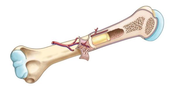
The outer part of a long bone is made of compact bone. The outer part of a long bone is made of compact bone. Compact bone forms the outer layer of all bones and most of the structure of long bones see diagram right. Sclerostin inhibits bone formation mostly by antagonizing lrp5/6, thus inhibiting wnt signaling. Human skeleton diagram unlabeled graph diagram.
2018 2019 Long Bone Anatomy Human Anatomy Quiz Quizizz from media.quizizz.com These units allow compact bone to. Femur bone diagram unlabeled via. Between the rings of matrix, the bone cells (osteocytes) are located in spaces called lacunae. Its unlabeled, so that your practce better. The bones shown in the chest and hip region in the labeled human skeleton diagram are the ribs, vertebrae, pelvis, os coxae, sacrum and coccyx. Location of red and yellow marrow in adults and. What are diplo , its function, and location? Human gross anatomy study | humandiagram.info.
Compact bone is the denser, stronger of the two types of osseous tissue (figure 6.3.6).
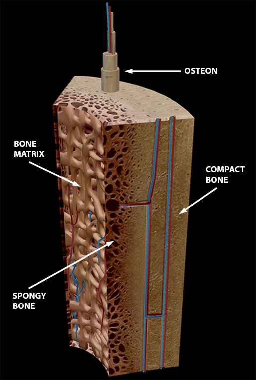
Key.' carotid canal coronal suture ethmoid bone external occipital protuberance foramen lacerum foramen magnum foramen ovale frontal bone edwnq'p'iep'n glabella. The osteon consists of a central canal called the osteonic (haversian) canal, which is surrounded by concentric rings (lamellae) of matrix. Unlabeled diagrams of the appendicular skeleton. Sclerostin inhibits bone formation mostly by antagonizing lrp5/6, thus inhibiting wnt signaling. Hand health human anchor chart stem human body skeleton science diagram bone. Bone classification, structure & relationships: Foot bone diagram unlabled wiring diagram t3. Compact bone is made of a matrix of hard mineral salts reinforced with tough collagen fibers. Compact bone, also known as cortical bone, is a denser material used to create much of the hard structure of the skeleton. Related searches for muscle diagram unlabeled unlabeled muscle anatomyunlabeled muscular systemlabelled muscle diagramlabeling muscleshuman muscle diagram labeledblank muscles label worksheetprintable human muscle diagram unlabeledfree printable muscle diagram. This lab is designed to provide students with an overview of bones through a variety of investigative to identify the major regions and structures of an osteon in a histological specimen of compact bone (or diagram or model of one). Total there are 12 pairs of ribs, as you can see in the diagram. (b) in this micrograph of the osteon, you can clearly see the concentric lamellae and central canals.
Muscles leg part posterior compartment. Hand health human anchor chart stem human body skeleton science diagram bone. Label compact and spongy bone illustrations as demonstrated in class. Sclerostin inhibits bone formation mostly by antagonizing lrp5/6, thus inhibiting wnt signaling. Students fill in the boxes with the names of the bones.
Compact Bone Tissue Model from www.elcamino.edu The bones mentioned in each human skeleton chart are: Between the rings of matrix, the bone cells (osteocytes) are located in spaces called lacunae. (b) in this micrograph of the osteon, you can clearly see the concentric lamellae and central canals. The osteon consists of a central canal called the osteonic (haversian) canal, which is surrounded by concentric rings (lamellae) of matrix. Students fill in the boxes with the names of the bones. 13 photos of the compact bone diagram labeled. Label compact and spongy bone illustrations as demonstrated in class. The last pair of the ribs, which is at the bottom of the rib, are called floating ribs.
What are diplo , its function, and location?
As seen in the image compact bone is formed from a number of osteons, which are circular units of bone material and blood vessels. It is a bone is one of two kinds of bone tissue that can be found in the compact type of bone wraps around and protects the only other type of bone tissue known as the you should include the histology of compact bone slides with diagram as well into your article. Related images with foot bone diagram unlabled. Minks bone diagram diagram base website bone diagram fishbonelabdiagramtemplate levantorosadeiventi it. Femur bone diagram unlabeled via. Unlabeled skeleton hand bone anatomy, anatomy bones, gross anatomy, medical. Hand, grasping organ at the end of the forelimb of certain vertebrates that exhibits great mobility and flexibility in the digits and in the whole organ. Compact bone consists of closely packed osteons or haversian systems. Unlabeled diagrams of the appendicular skeleton. Long bone structure diagram and definitions flashcards quizlet. The bones shown in the chest and hip region in the labeled human skeleton diagram are the ribs, vertebrae, pelvis, os coxae, sacrum and coccyx. Its unlabeled, so that your practce better. Compact bone, also known as cortical bone, is a denser material used to create much of the hard structure of the skeleton.
The bones shown in the chest and hip region in the labeled human skeleton diagram are the ribs, vertebrae, pelvis, os coxae, sacrum and coccyx. Muscles leg part posterior compartment. 13 photos of the compact bone diagram labeled. Human skeleton print cut outs | unlabeled human skeleton diagram. Anchor chart human bone diagram human body skeleton stem science health hand.
3d Skeletal System Compact Bone Spongy Bone And Osteons Oh My from www.visiblebody.com A typical long bone showing gross anatomical features. Human tongue anatomy vector image. Sclerostin inhibits bone formation mostly by antagonizing lrp5/6, thus inhibiting wnt signaling. Compact bone, also known as cortical bone, is a denser material used to create much of the hard structure of the skeleton. Appendicular skeleton quiz diagram clip art at clker com vector. Bone classification, structure & relationships: Printable animal cell diagram u2013 labeled unlabeled and blank. The osteon consists of a central canal called the osteonic (haversian) canal, which is surrounded by concentric rings (lamellae) of matrix.
Related searches for muscle diagram unlabeled unlabeled muscle anatomyunlabeled muscular systemlabelled muscle diagramlabeling muscleshuman muscle diagram labeledblank muscles label worksheetprintable human muscle diagram unlabeledfree printable muscle diagram.
Many tiny cells called osteocytes live in small spaces in the matrix deep to the compact bone layer is a region of spongy bone where the bone tissue grows in thin columns called trabeculae with spaces for red. Parts of a long bone unlabeled diagram system. Muscles leg part posterior compartment. Human tongue anatomy vector image. Compact bone consists of closely packed osteons or haversian systems. .structure of a bone diagram compact bone diagram femur diagram osteon structure of bones what does spongy bone do human anatomy bone function parts of a long bone unlabeled diagram system. Human gross anatomy study | humandiagram.info. Minks bone diagram diagram base website bone diagram fishbonelabdiagramtemplate levantorosadeiventi it. 13 photos of the compact bone diagram labeled. What are diplo , its function, and location? Location of red and yellow marrow in adults and. The outer part of a long bone is made of compact bone. Related images with foot bone diagram unlabled.
A typical long bone showing gross anatomical features compact bone diagram. Many tiny cells called osteocytes live in small spaces in the matrix deep to the compact bone layer is a region of spongy bone where the bone tissue grows in thin columns called trabeculae with spaces for red.


0 Komentar